Optical Coherence Tomography (OCT)
By obtaining Optical Coherence Tomography (OCT) images, we are able to view the distinct retinal layers in the eye. An eye care professional can use these images to diagnose, track, and treat certain ocular diseases.
When obtaining OCT images, the ophthalmic technician will ask the subject to sit comfortably facing forward. The subject will then place his/her chin on a chin rest and forehead against a forehead rest and will be asked to look at a blue light. The ophthalmic technician will then zoom in the camera very close to the eye; however the eye will not be touched by the equipment. The technician will obtain two different scans (and possibly more) of each eye. The first scan obtained will be a full scan of the macula (or central acuity region) of the eye.This scan takes about 30 seconds if the subject is able to focus only on the blue light, sometimes this scan takes longer if the subject has certain ocular conditions. The technician will then take several pictures of the optic nerve by asking the subject to again look at the blue light, now displaced from the original location. These pictures are quick and take around 10 seconds. Typical OCT images are shown below.
When obtaining OCT images, the ophthalmic technician will ask the subject to sit comfortably facing forward. The subject will then place his/her chin on a chin rest and forehead against a forehead rest and will be asked to look at a blue light. The ophthalmic technician will then zoom in the camera very close to the eye; however the eye will not be touched by the equipment. The technician will obtain two different scans (and possibly more) of each eye. The first scan obtained will be a full scan of the macula (or central acuity region) of the eye.This scan takes about 30 seconds if the subject is able to focus only on the blue light, sometimes this scan takes longer if the subject has certain ocular conditions. The technician will then take several pictures of the optic nerve by asking the subject to again look at the blue light, now displaced from the original location. These pictures are quick and take around 10 seconds. Typical OCT images are shown below.
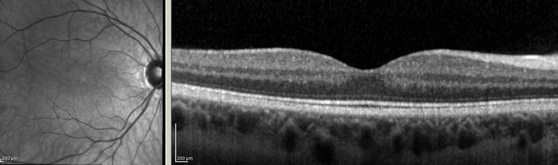
OCT images of a healthy adult subject. Left image: Full view of the retina; Right image: retinal layers and fovea.
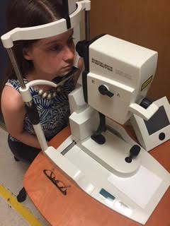
The OCT imaging machine.
References:
http://www.opsweb.org/?page=RetinalOCT\
http://www.heidelbergengineering.com/us/products/spectralis-models/imaging-modes/spectral-domain-oct/
http://www.opsweb.org/?page=RetinalOCT\
http://www.heidelbergengineering.com/us/products/spectralis-models/imaging-modes/spectral-domain-oct/