Fundus Photography
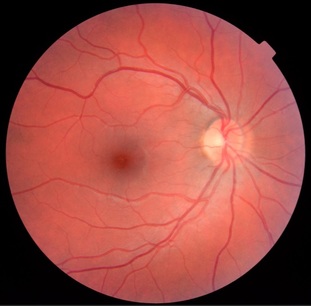
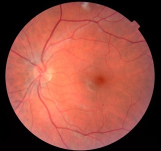
Fundus photography allows us to image the retina, the layer of the eye that is light-sensitive. The retina is red due to the large amount of blood that is supplied to the region. Typical images of the retina obtained using the Topcon camera are shown below. Eye care professionals are able to use these images to diagnose and monitor certain diseases.
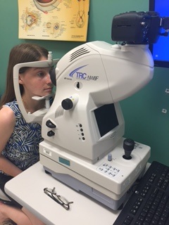
To obtain an image of the retina using the Topcon camera, subjects will be asked to place their chin on a chin rest and forehead on a forehead rest and to stare at a green light. The ophthalmic technician will then zoom in the camera very close to the eye; however the eye will not be touched by the equipment. When the technician is ready, he/she will give a short countdown and take the photo. There is a bright flash from the camera when the photo is taken. There are many regions of the retina that the photographer can take pictures of, thus the technician may ask the subject to look in other directions than where the green light is located and take additional pictures.
To obtain an image of the retina using the Topcon camera, subjects will be asked to place their chin on a chin rest and forehead on a forehead rest and to stare at a green light. The ophthalmic technician will then zoom in the camera very close to the eye; however the eye will not be touched by the equipment. When the technician is ready, he/she will give a short countdown and take the photo. There is a bright flash from the camera when the photo is taken. There are many regions of the retina that the photographer can take pictures of, thus the technician may ask the subject to look in other directions than where the green light is located and take additional pictures.
References:
http://www.opsweb.org/?page=fundusphotography
http://www.nlm.nih.gov/medlineplus/ency/article/002291.htm


