Multi-focal ERG (mfERG)
A specialized type of electroretinogram, called the multifocal ERG (mfERG), allows responses to be simultaneously recorded from multiple retinal areas using a stimulus array made up of black and white, flickering hexagons (a). The results can be analyzed to provide a contour map (b) of the function of the center of the retina, called the macula. This is the part of the eye that is used to read letters. It is also the last part of the retina to develop.
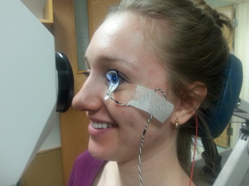
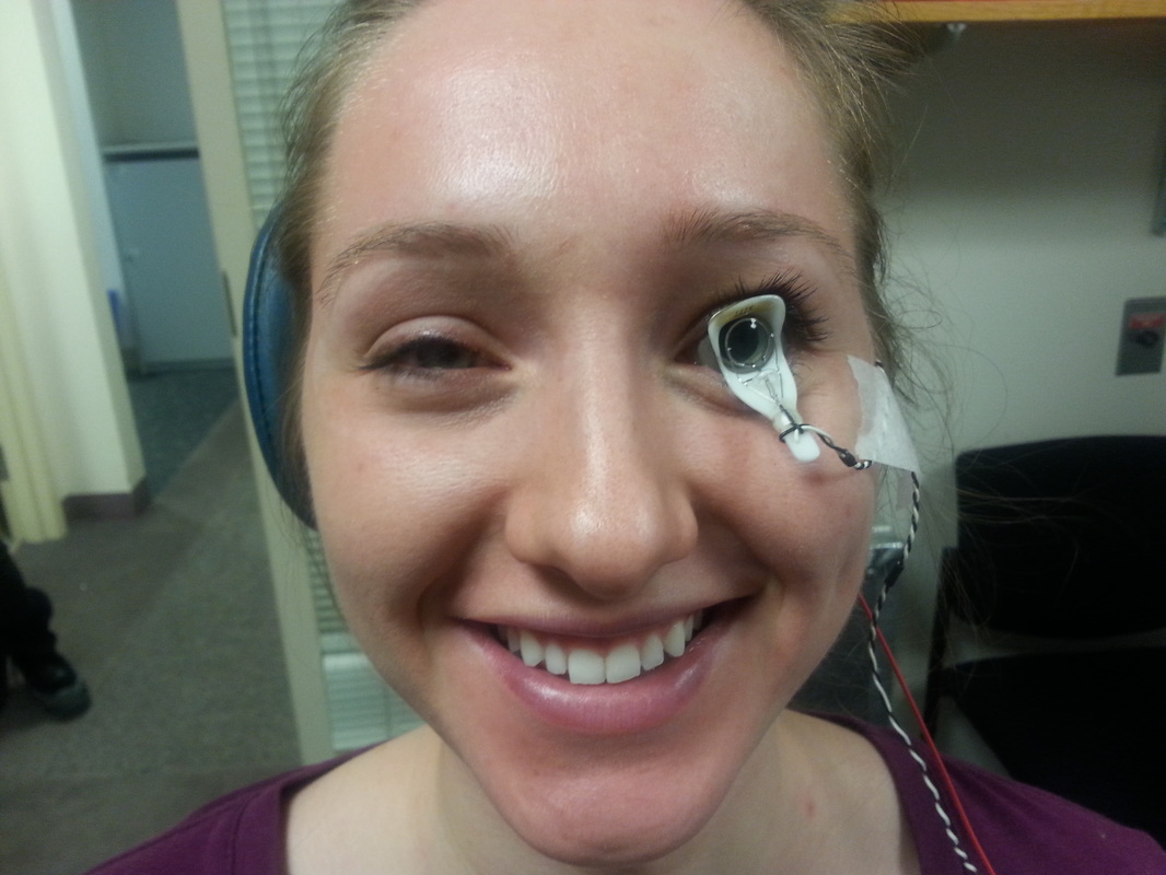
To prepare for the ERG test, drops are placed in the child's eyes to dilate the pupils. These drops are the same drops that eye doctors routinely use for eye examinations. They take about 30 minutes to take full effect. Next, anesthetic drops are given and a special contact lens is placed on the eye.
Infants and young children are positioned on the doctor's lap in front of a video monitor. Older children and adults sit alone. The contact lens picks up electrical signals produced by the retina, following which a map of the functional integrity of the retina is plotted.
The time for actual recording is typically 20-30 minutes; the total time (including dilation) is typically less than an hour.
A specialized type of electroretinogram, called the multifocal ERG (mfERG), allows responses to be simultaneously recorded from multiple retinal areas using a stimulus array made up of black and white, flickering hexagons (a). The results can be analyzed to provide a contour map (b) of the function of the center of the retina, called the macula. This is the part of the eye that is used to read letters. It is also the last part of the retina to develop.
To prepare for the ERG test, drops are placed in the child's eyes to dilate the pupils. These drops are the same drops that eye doctors routinely use for eye examinations. They take about 30 minutes to take full effect. Next, anesthetic drops are given and a special contact lens is placed on the eye.
Infants and young children are positioned on the doctor's lap in front of a video monitor. Older children and adults sit alone. The contact lens picks up electrical signals produced by the retina, following which a map of the functional integrity of the retina is plotted.
The time for actual recording is typically 20-30 minutes; the total time (including dilation) is typically less than an hour.
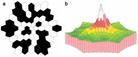
(a) The stimulus that the subject will be asked to look at during the mfERG.
(b) This image provides us with a "map" of a normal retina's response to the presented stimuli. The peak of the image represents the highest activity by the central retina.
|
|
One of our wonderful subjects completing the mfERG